Exploring Viruses in Augmented Reality: A New Perspective
Written on
Chapter 1: Introduction to Augmented Reality in Biology
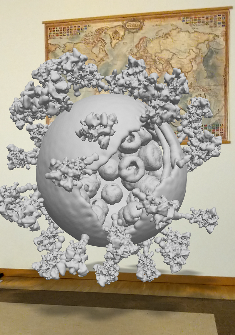
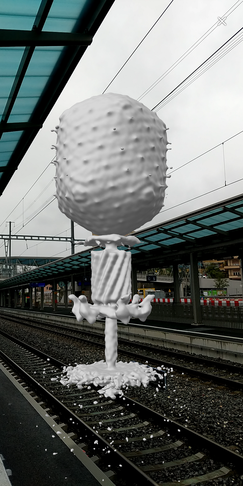
Augmented reality (AR) has revolutionized the way we visualize complex biological structures. Through the use of cutting-edge technology, we can now observe actual experimental reconstructions of viruses rather than mere illustrations. By utilizing the moleculARweb platform, individuals can effortlessly view these 3D models on their smartphones without the need for any app installation.
Here, I will guide you through various AR views of significant viral entities that are available for you to explore directly on your device.
Section 1.1: What Are the Models?
The models presented are not just simple sketches; they are sophisticated 3D reconstructions derived from high-resolution microscopy techniques, specifically cryoelectron tomography, rather than standard optical microscopy. These models are archived in the Electron Microscopy Data Base (EMDB), with raw data made accessible for validation. Below, I provide links to the original EMDB entries along with peer-reviewed publications for those interested in deeper insights.
Subsection 1.1.1: T4 Bacteriophage in AR

Section 1.2: P22 Bacteriophage's Intricate Structure

Chapter 2: The Impact of SARS-CoV-2

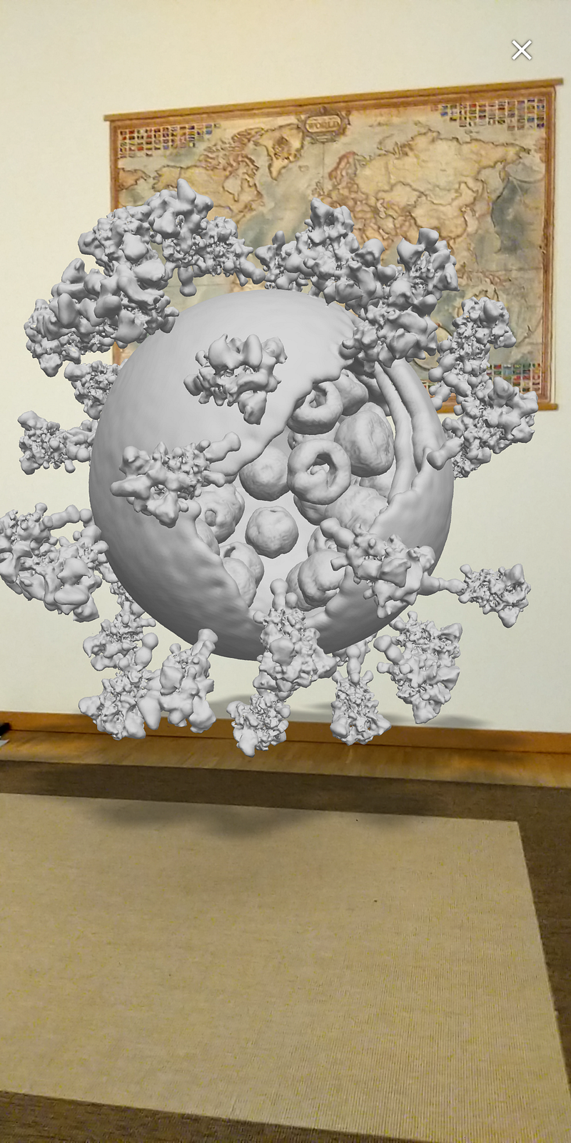
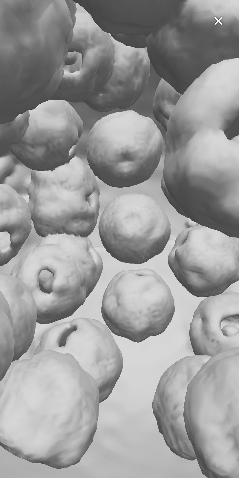
Within the virus, you'll find ribonucleoparticles, which contain the genetic material, as well as Spike proteins that facilitate infection by binding to the host's cell receptors.

I hope you find these AR views engaging and informative. For optimal results, use Chrome or Firefox on your smartphone. Should you encounter any difficulties, feel free to reach out for assistance. Rest assured, these models pose no risk of infection!
For those intrigued by web AR technology, I recommend checking out an article on two free AR tools designed for chemistry and biology education: